Article Outline
3. Performative, subjective and physiological indicators of alertness
4. Summary of the selected studies
Declaration of competing interest
Figures and tables
Volume 9 Issue 2 pp. 150-163 • doi: 10.15627/jd.2022.12
Alerting Effect of Light: A Review of Daytime Studies
Margarita Alwalidi,* Sabine Hoffmann
Author affiliations
Department of Civil Engineering, Technische Universität Kaiserslautern, Germany
*Corresponding author.
alwalidi@rhrk.uni-kl.de (M. Alwalidi)
sabine.Hoffmann@bauing.uni-kl.de (S. Hoffmann)
History: Received 4 May 2022 | Revised 16 July 2022 | Accepted 2 August 2022 | Published online 24 August 2022
Copyright: © 2022 The Author(s). Published by solarlits.com. This is an open access article under the CC BY license (http://creativecommons.org/licenses/by/4.0/).
Citation: Margarita Alwalidi, Sabine Hoffmann, Alerting Effect of Light: A Review of Daytime Studies, Journal of Daylighting 9 (2022) 150-163. https://dx.doi.org/10.15627/jd.2022.12
Figures and tables
Abstract
Light affects humans beyond only image formation. Several studies have reported that light can increase daytime alertness and can therefore be positively utilized to counter daytime fatigue and increase productivity in workspaces. This systematic review summarized and analysed relevant literature that investigated the daytime alerting effect of light. Using keywords “alertness” and “light” in the title we retrieved a total of 142 studies via three search engines. Out of 142 studies, only 26 investigated the alerting effect of light during daytime. Six studies were excluded from the review based on the exclusion criteria. We reviewed twenty articles from the year 1991 until 2022 that have investigated the alerting effect of polychromatic and monochromatic light sources. Only seven out of twenty studies found an alerting effect that was recorded through positive self-assessment or enhancement of cognitive processes. The studies in which the lighting application would be transferrable for office use were highlighted. A specific common trend regarding the enhancement of alertness was not detected, as different lighting conditions and protocols were applied in the reviewed studies. More studies that investigate the effect of polychromatic light with intensities and spectral compositions that are suitable for application in realistic working environments are necessary to determine whether the daytime alerting effect of light will be significant.
Keywords
Daytime, Alertness, Non-visual photoreception, Lighting
1. Introduction
1.1. Background
Office workers spend a large portion of their time in an indoor setting. Given the length of time that people spend at work, it is critical to create an environment that promotes their health and overall wellbeing. As of today, specifications for light vary based on the purpose of space but are limited to requirements for illuminance, glare and colour-rendering index [1]. Meanwhile, the desired outcome for office spaces is primarily focused on enhancing office workers' daytime productivity through the provision of favourable working environment including optimal illumination, being that light can impact the performance of people beyond its photopic impacts [2,3].
Recent discoveries in the field of biology indicate that light can inform visual as well as non-visual optical systems [4]. In light of this finding, it is no longer sufficient to evaluate light only using photopic metrics that do not account for non-visual effects. However, a metric that accurately represents both types of photoreception is yet to be determined. The concept of non-image-forming photoreception in humans is relatively new and is linked to the discovery of melanopsin containing intrinsically photosensitive retinal ganglion cells (ipRGCs) [2,5]. In recent years, research has provided ample support for the assertion that ipRGCs play a major role in the light-induced resetting of the circadian clock and melatonin suppression [6]. There is a rapidly growing body of literature, which suggests that light affects alertness, cognitive performance and physiological responses in humans [2,7–9].
As of now, many experimental studies have been done in an attempt to extend current knowledge on the non-visual effects of light on human health. However, the reports are inconsistent, especially in studies that investigate the non-visual effects of light during daytime. According to several publications, bright light [10–16] or light with high correlated color temperature (CCT) [17–19] has been shown to improve subjective ratings, performance and physiological indicators of alertness. Whereas, certain studies report no alerting effect of light at all [20–22]. The difference in results may be attributed to different protocols that were implemented in the studies, such as different exposure durations, intensity and spectral power distribution of light sources along with many other possible variations.
The aim of this literature review is to systematically present the protocols and the results of each study that investigates the daytime alerting effect of light. We have selected twenty studies and presented the findings of the review with consideration of possible implications for the built environment.
1.2. Visual and non-visual photoreception
In mammals, light is processed through visual and non-visual systems. Cones and rods are photoreceptors that mediate image-forming processes; they are responsible for photopic and scotopic visual functions respectively. Cones fall into three categories (S-, M-, L-cones) based on the type of photopigment they contain, which distinguishes their spectral sensitivity (see Fig. 1). Similarly, rods contain rhodopsin photopigment that has the maximum spectral sensitivity at ~ 505 nm.
Figure 1
![Relative spectral sensitivity curves of five photoreceptors with the respective photopigments [35].](figures/9-150-1.jpg)
Fig. 1. Relative spectral sensitivity curves of five photoreceptors with the respective photopigments [35].
Recently discovered ipRGC photoreceptors contain melanopsin, a photopigment with the peak spectral sensitivity in the short-wavelength range (~ 480 nm) [4,23–25]. The ipRGCs transmit light information to the suprachiasmatic nuclei (SCN), which is regarded as the mammalian internal clock. This prompts SCN to synchronize physiological processes to fit a near-24-hour circadian cycle [26]. The circadian cycle coordinates a number of mechanisms that influence the feeling of sleepiness, alertness, vigilant attention, psychomotor and perceptual-cognitive speed, and working memory [27–29]. Light can effectively alter the circadian rhythm and plays an important role in mediating alerting effects [30]. Lockley et al. have determined that timing, intensity, duration, pattern, wavelength and previous history of light exposure have a resetting effect on the circadian rhythm [30].
In well-sighted mammals, non-image-forming (NIF) responses to light are partially sustained by signals from cones in addition to the dominant inputs from melanopsin-expressing photoreceptors [31,32]. Yet, experimental studies indicate that even some blind individuals with preserved ipRGCs can experience melatonin suppression [33], reset of the circadian pacemaker and enhanced alertness when exposed to cool-white or short-wavelength light despite non-functioning cones and rods [34].
1.3. Non-visual effects of light in the built environment
Light is an important part of the built environment. Current regulations rarely consider light beyond its visual effect. As of today, there are just a few metrics that evaluate light non-photopically, such as the circadian stimulus (CS), circadian action factor (acv) and the equivalent melanopic lux (EML).
Circadian stimulus (CS) is a metric that considers the interaction of all photoreceptors and indicates the percentage of melatonin suppression after one hour of exposure to light. CS of 0.7 indicates saturation or 70 % of melatonin suppression. The authors of this metric report that CS of 0.3 in the early part of the day stimulates the circadian system and is associated with better sleep and improved behaviour and mood [36] even though it is not clear whether the effect of light is sustained beyond one hour of the exposure.
The circadian action factor (acv) is another metric that describes the circadian efficiency of light sources. It is the resulting ratio of the irradiance of any light source that is weighted by the circadian and the photopic spectral sensitivity functions. However, to our best knowledge, no recommendation for this ratio is proposed in the literature.
EML is a metric that weights the spectral power distribution of light with the spectral sensitivity function of ipRGCs instead of the photopic function V(λ). WELL Building Standard suggests at least 250 EML in work areas during daytime and no more than 50 EML in living areas in the evening to support circadian health [37].
Recently a preprint with recommendations for healthy daytime, evening and nighttime light exposure was published [38]. The authors of the preprint recommend exposure to a minimum of 250 EML at the eye level during daytime. They recommend exposure to daylight but also deemed polychromatic white light as an acceptable option. For evening time, 10 EML at the eye level should not be exceeded. During the nighttime, the light level should not exceed 1 EML at eye level. For nighttime activities that are unavoidable 10 EML at the eye level is an acceptable degree of exposure.
However, no single action spectrum can predict the NIF responses. Because of this, the International Commission of Illumination (CIE) has adopted five α-opic metrics that weight the spectral power distribution of light with the sensitivity function of the five photoreceptors (Fig. 1) in the eye [39]. Despite this, the standard does not provide recommendations regarding the appropriate magnitude of the α-opic metrics.
Since the discovery of the ipRGC photoreceptor is relatively recent, there are no definitive recommendations concerning the non-image-forming effects of light, although many studies reveal that light can counter sleepiness at night [40–42]. It is generally understood that exposure to shortwaves should be avoided before bedtime to prevent cognitive arousal.
On the other hand, studies also report that light can increase morning vitality [43] and counter daytime mental fatigue [44]. If in fact so, then it is of prime interest to investigate the effectiveness of light in enhancing alertness especially if it can be translated into sustained attention and increased productivity during daytime.
1. Research protocol
2.1. Purpose
The purpose of this review is twofold: if the results of the studies illustrate the same trend, one could utilize this information to increase daytime alertness in office spaces or other environments. On the other hand, if no uniform conclusion can be derived from the studies, this literature review will highlight, which studies present the most relevant framework with regard to realistic workspaces or other environments for further investigations.
2.2. Objectives
The objective of this study is to identify studies that investigate alerting effects of light during daytime, systematically summarize them and analyse whether the results show the same trend. If there is a definite tendency showing that people become more alert, then the aim is to identify which characteristics of a light source cause the influence and what time of day these effects are the most evident. In the case of a lack of such findings, a suggestion based on the identified gap in the studies should be proposed.
2.3. Criteria for including studies in the review
The studies for the review satisfied the inclusion criteria if the following requirements were met:
- Keywords “alertness” and “light” were both found in the title.
- The publications were original research.
- The experiments took place during daytime.
- The light condition was mediated by daylight or an electrical light source.
- Properties of the light source are reported and can be reproduced (i.e. documented intensity, spectral characteristics, light transitions).
- No administration of external influencing factors such as exogenous melatonin.
2.4. Search methods
The search for the relevant literature was carried out between March of 2021 and July 2022 via Science Direct, PubMed and Scopus. The engine searches returned 142 publications in total, 109 were original research papers. Duplicated studies across the three search engines were removed leaving 65 papers to review. Of those 65 papers, only 26 dealt with the daytime alerting effect of light. Six studies were eliminated based on their unsuitability (Fig. 2). Twenty studies that were left to review were split into those that used polychromatic (n=16) or monochromatic light (n = 4) sources. Studies that utilized polychromatic light sources were further subdivided into those that investigated the effect of illuminance (n = 10) and correlated colour temperature (n = 6).
Figure 2
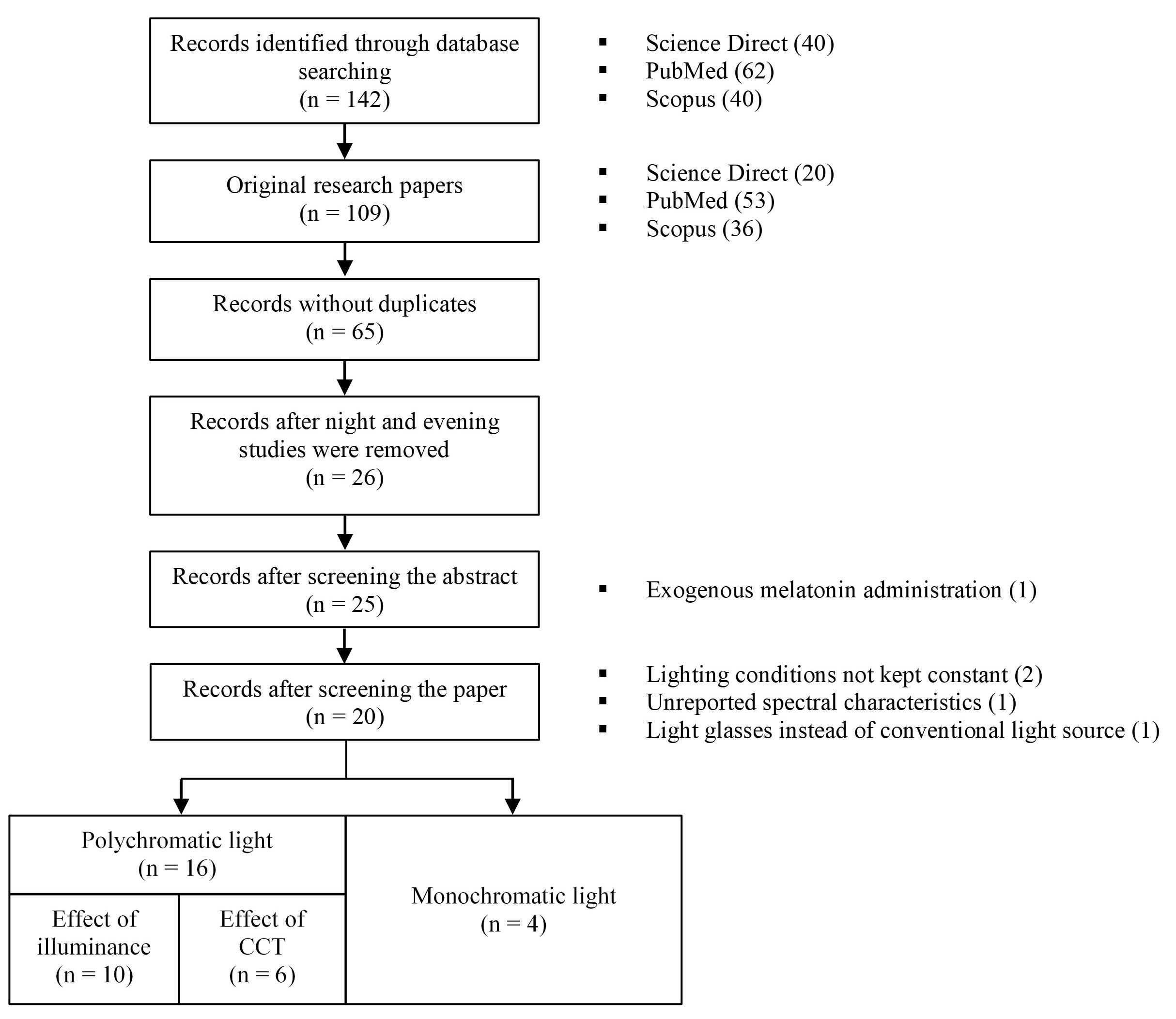
Fig. 2. Summary of the selection process for the review.
2.5. Selected studies
The publication date of the selected studies ranged from 1991 until 2022. The experiments differ in their setup and population sizes. The minimum age of the test subjects was 18 years and the maximum age was 65 across all the studies. The location and the date of the experiments were not reported in every study, even though the season is significant for such assessments, as the effect of light during months with limited daylight has been linked to more pronounced effects [13,45]. Despite this, these studies were retained for the analysis, due to a relatively low number of studies that investigate the alerting effect of light during daytime.
Tables 1-3 present the selected studies and the performative, subjective and physiological indicators of alertness utilized by the respective authors (see section 3). Table 1 summarizes the studies in which the authors compared the illuminance of polychromatic light. Table 2 presents the studies with polychromatic light and variable CCT (and illuminance in some cases). Table 3 shows the studies in which authors used monochromatic light.
Table 1
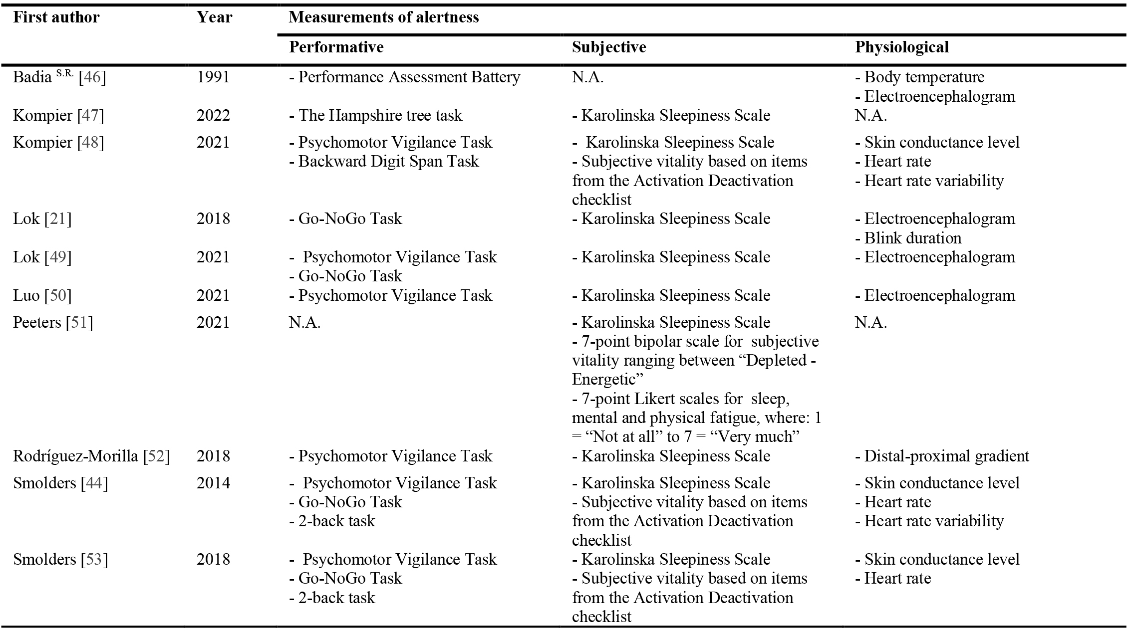
Table 1. Studies that investigate the impact of illuminance. Measurements of alertness: performative, subjective and physiological indicators.
Table 2
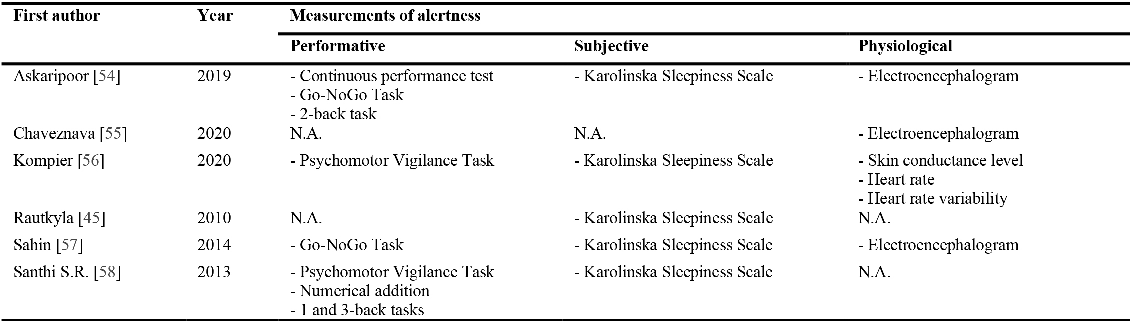
Table 2. Studies that investigate the impact of CCT. Measurements of alertness: performative, subjective and physiological indicators.
Table 3
Table 3. Studies that investigate the impact of monochromatic light. Measurements of alertness: performative, subjective and physiological indicators.
3. Performative, subjective and physiological indicators of alertness
The effect of light is usually evaluated based on performative, subjective and physiological indicators in such studies. Physiological indicators can sometimes correlate with changes in subjective alertness or cognitive performance. These indicators are often clinically assessed by tracking physiological responses to light stimuli. For example, melatonin concentration is a common biomarker used for monitoring circadian rhythm. A high level of melatonin promotes sleep and is expected at night [63]. Some studies report a partial causal role of melatonin suppression in mediating the alerting effects of light [64,65]. Melatonin is not produced during the day, therefore it is not used as a physiological indicator of elevated alertness in none of the selected studies.
Daily fluctuations in the core body temperature are regarded as markers of the circadian clock. It has been reported that increased body temperature is correlated with improved performance and alertness [66]. Another physiological indicator is an increased distal-proximal skin temperature gradient (i.e., a bigger temperature difference between distal and proximal temperature), which has been previously linked to a decrease in sleepiness [67].
Another commonly measured response to light manipulations is the skin conductance level, which determines the electrical conductivity of the skin. Measuring skin conductance level reflects the psychological or physiological arousal in response to stimuli.
Electroencephalography (EEG) monitors electrical brain activity, otherwise referred to as brain waves. The amplitude of the brain signals is very small but can be picked up by EEG. The signals detected by EEG are classified into five main types, depending on their frequency (listed in descending order): gamma, beta, alpha, theta and delta [68]. In general, frequencies below (and including) alpha are more sensitive to changes in vigilance as opposed to gamma and beta waves [69]. The increase in theta and delta activity leads to fatigue [70]. Baek et al. [71] hypothesized that the decrease of activity in lower and upper alpha frequencies is associated with increased attentional processing and semantic memory performance respectively. This method of monitoring was used in many of the selected studies.
Additionally, heart rate and heart rate variability are used to trace physiological changes in response to manipulations with light. According to research findings, heart rate and heart rate variability reflects vigilant attention in people who are subjected to partial chronic sleep restriction [72].
To confirm that physiological changes translate to the improvement of alertness, experimental studies usually include various performance-measuring tests and subjective self-assessment questionnaires. Of the most commonly used objective performance tests is the Psychomotor Vigilance Task (PVT) [73]. It measures sustained alertness and vigilance by measuring the reaction time of response to stimuli while also recording the number of attentional lapses [74]. However, the Continuous Performance Test (sustained and selective attention) [75], N-back task (memory) [76], the Stroop task (processing speed) [77] and Go-NoGo Task (sustained attention and response control) [78] are also often used in such studies.
Subjective self-assessment questionnaires provide valuable information about the personal perception of test subjects. They are uncomplicated to conduct and suitable for large groups. Karolinska Sleepiness Scale (KSS) [79] is the most commonly used questionnaire that measures subjective sleepiness and alertness. Stanford Sleepiness Scale (subjective sleepiness) [80] and Activation-Deactivation Adjective Check List (energetic arousal and tenseness) [81] along with other self-assessment questionnaires are used to report the subjective perception of the participants.
4. Summary of the selected studies
4.1. Effect of the illuminance
Table 4 shows an overview of the publications that investigate the effect of the illuminance of polychromatic white light on alertness. The population size for studies ranged between 8 and 50. Badia et al. [46], Lok et al. [21] and Peeters et al. [51] recorded no increase in alertness neither in the morning nor in the afternoon when the illuminance of polychromatic white light was increased in comparison to respective control conditions. Similarly, Smolders et al. [53] reported only a very modest linear relationship between the log-transformed illuminance and subjective correlates of alertness.
Table 4
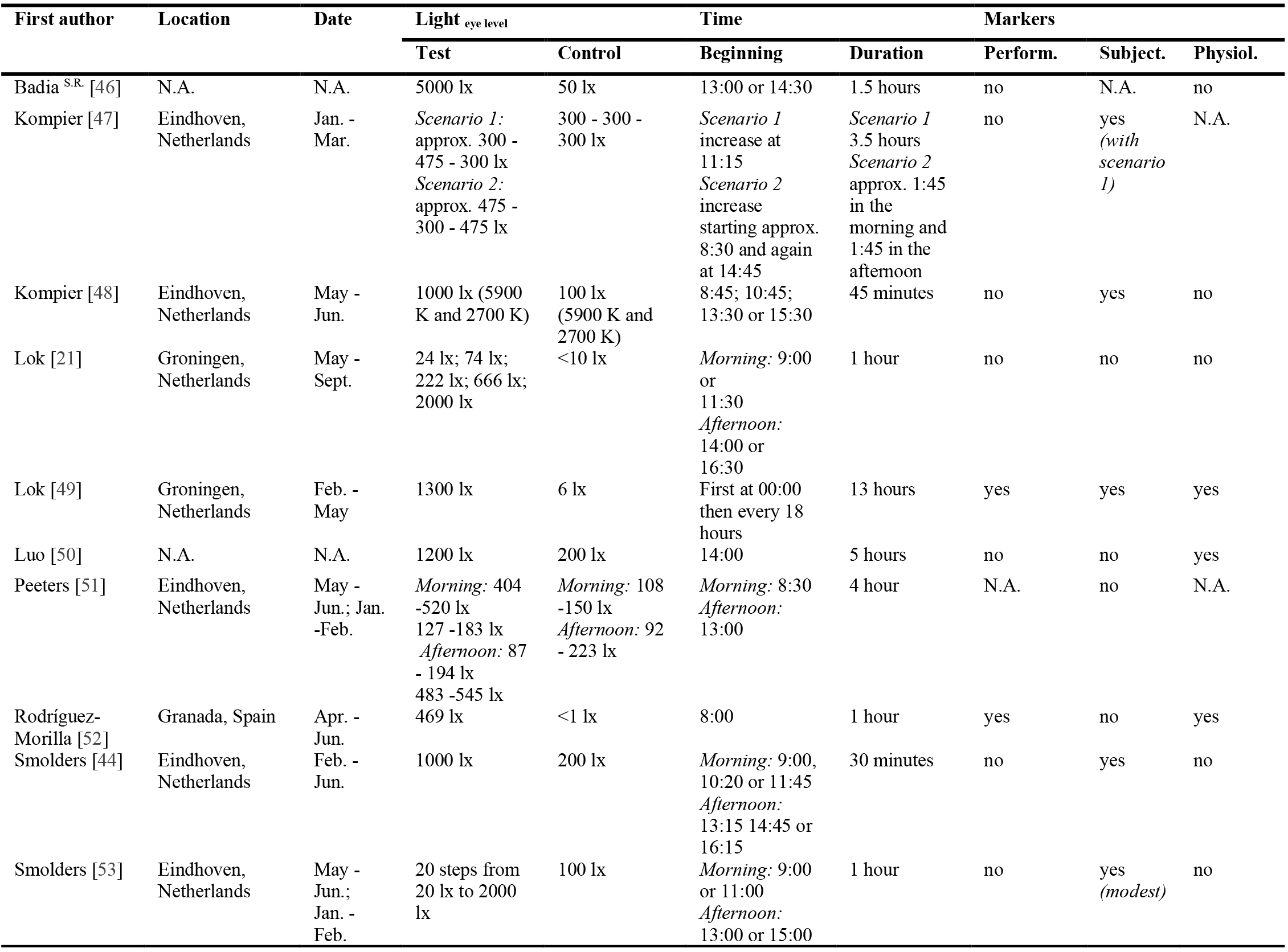
Table 4. Overview of the publications that investigate the impact of illuminance. S.R. = sleep restriction.
On the other hand, Kompier et al. [47] reported a modest reduction in subjective sleepiness when test subjects were exposed to bright light that was a combination of daylight and electrical light at noon. It is noteworthy, that it was the only study that investigated the impact of daylight in combination with electrical light.
In another study, Kompier et al. [48] concluded that an increase in subjective vitality and decrease in subjective sleepiness were attributed to the increase of illuminance and not CCT. They found the alerting effect to be immediate, as it lasted for at least 20 minutes and did not exceed the duration of the exposure.
A more recent study by Lok et al. [49] reported that bright light increased performative, subjective and physiological markers of alertness in comparison to dim light (< 6 lx) for 13-hour-long light exposure. However, in this study, the sleep of the test subjects was restricted. Meaning that the same results reported by this study cannot be expected for non-sleep restricted persons.
Luo et al. [50] compared the alerting effect of regular office light (1200 lx vs. 200 lx) for 5 hours in the afternoon and found that in comparison to subjective and performative indicators, the EEG activity may be more responsive to the bright light-induced alertness response during the day.
Rodríguez-Morilla et al. [52] reported that an hour-long, morning exposure to a blue-enriched polychromatic white light improved the performance as indicated by faster reaction time in performance-measuring tests and decreased distal-proximal skin temperature gradient of evening chronotypes in comparison to a dim light control condition (< 1 lx). However, Rodríguez-Morilla et al. reported no improvement in subjective sleepiness.
Smolders et al. [44] investigated the alerting potential of polychromatic white light at 1000 lx and 200 lx in countering mental fatigue. The duration of exposure was 30 minutes and took place in the morning and the afternoon. Based on their findings, the exposure with 1000 lx increased vitality and reduced subjective sleepiness in comparison to 200 lx. Nevertheless, the bright light exposure did not affect either performative or physiological markers of alertness.
4.2. Effect of the correlated colour temperature
Table 5 shows an overview of the publications that investigate the effect of the CCT of polychromatic white light (with monochromatic pulses [55]) on alertness. The population size was between 11 and 138 across the studies. All studies presented in this category recorded at the very least a minor effect of correlated colour temperature on alertness.
Table 5
![Overview of the publications that investigate the impact of correlated color temperature (with monochromatic pulses [55]).](figures/9-150-13.jpg)
Table 5. Overview of the publications that investigate the impact of correlated color temperature (with monochromatic pulses [55]).
Askaripoor et al. [54] suggest that exposure to light with 12000 K and 2700 K at 500 lx for 2 hours in the afternoon can improve the physiological correlates of alertness in EEG measurements in comparison to normal white light (4000 K at 500 lx) and dim light (3520 K at < 5 lx). However, the physiological changes did not translate to improvements in the performance of subjective alertness.
Chaveznava et al. [55] reported that the beta waves exhibited a statistical significance in arousal levels in EEG measurements as a result of exposure to blue and green LED pulses with dimly-lit background illumination for approximately one hour in the morning. Furthermore, the pupil reacted with larger constriction in this condition. The study did not examine the effects of light on performative and subjective markers of alertness.
Kompier et al. [56] have investigated the impact of light transitions on measures of alertness. The study took place during work hours for one and a half hours and the transition took place after 45 minutes. On average, participants felt significantly more vital and alert after the transition from warm to cool white light. Physiological indicators showed only minor changes. The skin conductance level was significantly lower in the transition from cool white light to warm white light compared to the constant cool white condition, signifying that the warm white light is less physiologically arousing for people.
Rautkyla et al. [45] investigated the effect of light with 3870 K and 12370 K on subjective alertness during lectures in autumn (1000 lx) and spring (800 lx). The autumn study reported that the exposure to a light source with 12370 K assisted students in maintaining higher levels of alertness in comparison to the 3870 K light in the afternoon. The authors reported no change in subjective alertness during the lecture period in spring.
Sahin et al. [57] compared the alerting effect of monochromatic red (213 lx) and polychromatic white (361 lx) light of the same irradiance (1.1 W/m²) with a dim (< 5 lx) light. The results demonstrated that red monochromatic light can reduce reaction time in the performance tests during a daytime two-hour long exposure. The alerting effect of red light was further confirmed via EEG by a significant decrease in alpha power in the middle of the afternoon.
Santhi et al. [58] studied whether sleep inertia exhibits an intensity and spectrum-dependent sensitivity in 4-hour-long light exposure in sleep-restricted subjects. Based on their findings, the light with 7717 K at 195 lx only improved the response speed on a performance test. Nevertheless, the clinical significance or practical relevance is not apparent because the performance improvement was small. Furthermore, 7280 K at 750 lx did not affect subjective sleepiness and most of the performance measures.
4.3. Effect of the monochromatic light
Table 6 shows an overview of the publications that investigate the effect of monochromatic light exposure on alertness. The population size was between 10 and 60 across the studies. Out of the four articles, the study by Segal et al. [60] has not recorded any alerting effect of exposure to blue, green or red monochromatic light.
Table 6
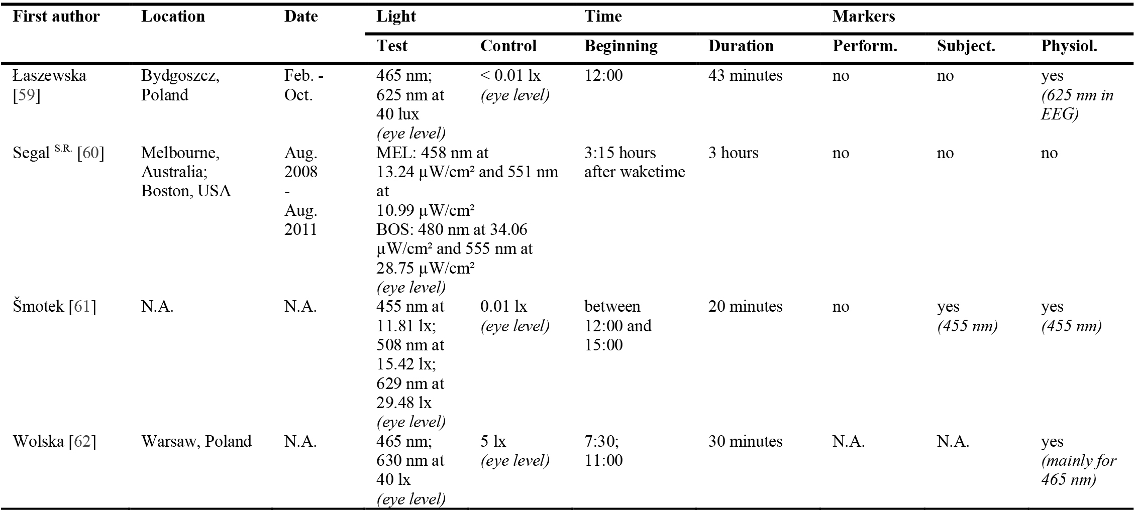
Table 6. Overview of the publications that investigate the impact of monochromatic light.
Łaszewska et al. [59] investigated whether exposure to red (625 nm) or blue light (465 nm) at 40 lx at noon would increase performative, subjective and physiological indicators of alertness. Exposure to red light reduced alpha power over time, however, in a small range. The authors have concluded that the alerting effect of light at noon is very limited and did not translate into performative or subjective improvements in alertness.
Šmotek et al. [61] compared the alerting effects of 455 nm, 508 nm and 629 nm at identical irradiance (14 µW/cm²) but different illuminance (Table 3) against dim light. The investigation took place in the afternoon and lasted 20 minutes. The authors concluded that the subjects felt less sleepy under light with 455 nm than under 508 nm, 629 nm or dim light. Furthermore, blue light enhanced cortical activity related to cognitive tasks as registered by EEG.
Wolska et al. [62] compared monochromatic light with 465 nm and 630 nm at 40 lx with dim light for 30 minutes in the morning. The study investigated only the physiological changes based on the EEG registration. According to their findings, there was a visible impact of blue light exposure on the increase in acute alertness level after the exposure as signified by theta and alpha bands. The impact was not apparent with red light, but the tendency of increasing the alertness was evident.
5. Discussion
Based on the summaries from section 3, only three [21,46,51] out of ten studies that investigated the effect of illuminance on alertness reported no changes in the performative, subjective and physiological indicators for alertness. It is noteworthy that two of the aforementioned studies utilized dim light in the control conditions (50 lx and <10 lx), even though people are commonly exposed to much higher illuminances during daytime. For this reason, even despite the discovery of subjective, performative and physiological changes in two other studies [49,52] the illumination conditions (6 lx and < 1 lx) of the experiments are not representative of the conditions in real offices and do not depict practical relevance in the framework of this review.
Another study reported that the increase in illuminance from 200 lx to 1200 lx for five hours starting at 14:00 elevated physiological markers of alertness. However, it did not enhance either subjective or performative indicators of alertness [50]. Nevertheless, if the physiological markers of alertness do not translate to either subjective or performative indicators then such a scenario would be ineffective for non-visual stimulation in an office setting.
Smolders et al. [53] have reported a linear relationship between the log-transformed illuminance and subjective correlates of alertness, however, the relationship was very modest. In another study by Smolders et al. [44], the subjects felt significantly less sleepy and more vital when they were exposed to polychromatic light with 1000 lx in comparison to 200 lx when the participants were suffering from mental fatigue.
In another study, Kompier et al. [48] found an immediate increase in subjective alertness when the subjects were exposed to 45 minutes of 1000 lx in comparison to 100 lx. The effect was temporary and lasted approximately 20 minutes. In a study with a dynamic lighting scenario, Kompier et al. [47] reported an increase in subjective alertness with even lower illuminance. They reported that the subjective sleepiness decreased when the illuminance increased from 300 lx to 475 lux for 3.5 hours around noon.
Figure 3 shows a summary of the reviewed studies that investigated the alerting effect of light through manipulation of illuminance. Figure 4 presents the range of illuminance that was used in the studies. The opaque bars present the studies in which an alerting effect was found.
Figure 3
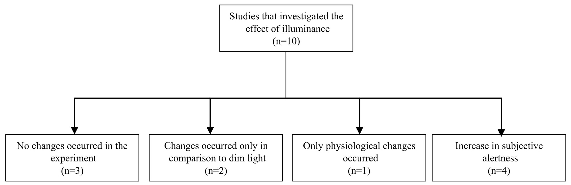
Fig. 3. Studies that investigated the effect of illuminance on alertness and the main conclusions.
Figure 4
![Studies that investigated the effect of illuminance on alertness. Opaque bars indicate studies that reported alerting effect (as long as it was not only a physiological and the control condition was not a dim light). A study by Peeters et al. [51] was presented in the figure four times: morning (AM) exposure with high and low illuminance and afternoon (PM) exposure with high and low illuminance.](figures/9-150-4.jpg)
Fig. 4. Studies that investigated the effect of illuminance on alertness. Opaque bars indicate studies that reported alerting effect (as long as it was not only a physiological and the control condition was not a dim light). A study by Peeters et al. [51] was presented in the figure four times: morning (AM) exposure with high and low illuminance and afternoon (PM) exposure with high and low illuminance.
Another six studies investigated the impact of the correlated colour temperature (CCT) on alertness (Figs. 5 and 6). One of the studies [55] only investigated the physiological markers of alertness. The authors reported the effect of blue and green LED pulses with dimly-lit background on the EEG correlates of alertness and pupil constriction, however, the study did not investigate if the physiological indicators of alertness would cause subjective or performative improvements.
Figure 5

Fig. 5. Studies that investigated the effect of CCT on alertness and the main conclusions.
Figure 6
![Studies that investigated the effect of CCT on alertness. Opaque bars indicate studies that reported alerting effect (as long as it was not only physiological and the control condition was not a dim light). Studies by Kompier et al. [56], Rautkyla et al. [45] and Santhi et al. [58] compare different scenarios in relation to each other, not to control light.](figures/9-150-6.jpg)
Fig. 6. Studies that investigated the effect of CCT on alertness. Opaque bars indicate studies that reported alerting effect (as long as it was not only physiological and the control condition was not a dim light). Studies by Kompier et al. [56], Rautkyla et al. [45] and Santhi et al. [58] compare different scenarios in relation to each other, not to control light.
Another group [54] reported that exposure to light with 12000 K and 2700 K at 500 lx can improve the physiological correlates of alertness in EEG measurements in the afternoon exposure. However, the physiological changes did not translate to performative and subjective improvements.
When a dynamic light was studied [56] it was concluded that a transition from low-intensity warm white light (2708 K) to high-intensity cold white light (5854 K) after 45 minutes can improve some of the physiological indicators as well as the feeling of alertness and vitality.
One of the studies [45] investigated the effect of warm (3870 K) and cool (12370 K) white light on the subjective feeling of sleepiness. They concluded that the alerting effect of cold white light is season-dependent and is pronounced in autumn.
Another study [57] compared monochromatic red light (631 nm) with polychromatic warm white light (2568 K). The study suggested that monochromatic red light can improve performance by reducing the reaction time in performance-measuring tests and some of the physiological markers. However, it did not improve the subjective feeling of alertness in subjects of the experiment.
Finally, the last study in this category [58] found that light with 7717 K at 195 lx improved the response speed on the performance test. Although, in the same study, the authors found no effect of 7280 K at 750 lx on subjective sleepiness and most of the performance measures. Furthermore, the sleep of the test subjects in this study was limited. Thereby, it is not clear if well-rested individuals would similarly respond to identical light.
The four studies from the last category investigated the impact of monochromatic light (Figs. 7 and 8). One of the studies [60] found no alerting effect of monochromatic blue (458 nm and 480 nm) and green (555 nm and 551 nm) light on performative, subjective and physiological indicators.
Figure 7
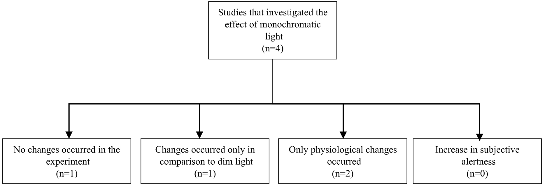
Fig. 7. Studies that investigated the effect of monochromatic light on alertness and the main conclusions.
Figure 8
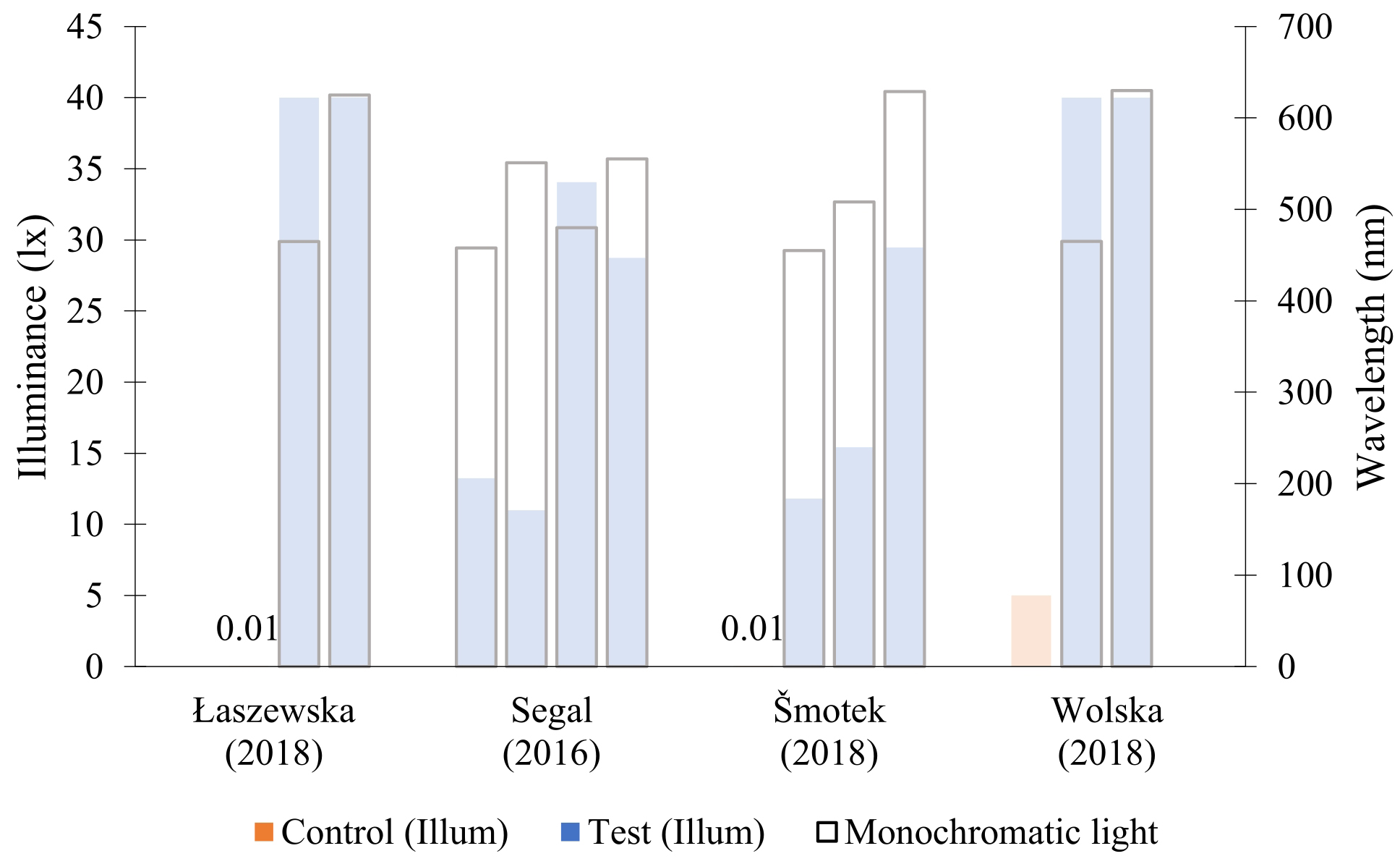
Fig. 8. Studies that investigated the effect of monochromatic light on alertness. Opaque bars indicate studies that reported alerting effect (as long as it was not only a physiological and the control condition was not a dim light). A study by Segal et al. compares different scenarios in relation to each other, not to control light. A study by Segal et al. reported the intensity of light as irradiance (µW/cm²) instead of illuminance.
Two of the studies [59,62] compared the alerting effect of monochromatic blue light (465 nm) with monochromatic red light (625 nm or 630 nm). Both studies reported an increase in the physiological markers of alertness, however with different conclusions. One study [59] reported that red monochromatic light enhances alertness measures in the EEG. The other study [62] concluded the same effect for the blue monochromatic light, although it was noted that the tendency of increasing the alertness with red monochromatic light was visible.
The last study [61] found that besides the physiological effect that was recorded by EEG, the sleepiness tests indicated that the participants were less sleepy under blue (455 nm) than under green (508 nm) or red (629 nm) light. However, these findings were established in comparison to a dim control condition (0.01 lx). Further investigation is necessary if the alerting effect would persist in comparison to a brighter control condition that is typical for an office environment.
It is noteworthy that monochromatic light is unsuitable for office illumination, therefore the results of the studies from this category cannot be directly translated into practice. It would be relevant to see if the same intensity of blue (455 nm) monochromatic light that was investigated in [61] would provide a physiological and subjective alerting effect within the spectrum of polychromatic white light. Additionally, the studies implemented very dim light as a control condition, even though people are usually experiencing significantly higher illuminance during daytime. Whether the alerting effect of monochromatic light is noteworthy in comparison to control conditions with higher illuminances remains to be determined.
Based on the results of the summary seven out of twenty studies concluded that light definitively enhances alertness by increasing performance or subjective indicators (illuminance-based studies: [47,48,44,53]; CCT-based studies: [56,45,57]). Table 7 shows the studies that recorded an alerting effect of light (beyond only the physiological effect).
Table 7
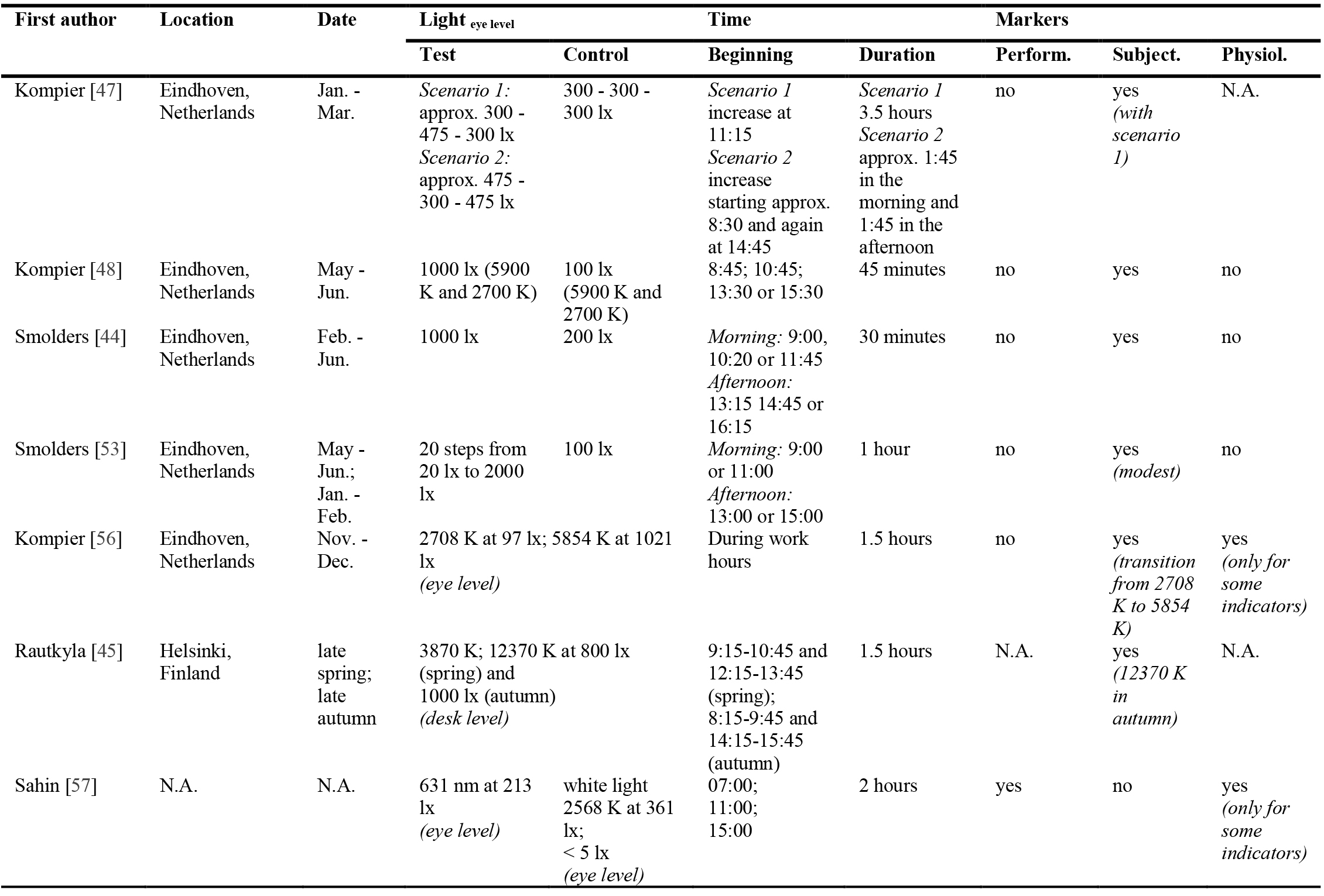
Table 7. Overview of the publications that reported alerting effect of light.
Based on this review, no uniform conclusion can be derived, as the alerting effect was achieved through different lighting manipulations in the studies. However, it can be concluded that more daytime studies that focus on the manipulation of CCT and the illuminance of polychromatic light are necessary. Additionally, to understand the non-visual implication of light in office spaces, the control condition should be brighter than dim light and comparable to what is commonly implemented in practice.
6. Conclusion
The concept of non-image-forming photoreception in humans is predominately mediated by melanopsin containing intrinsically photosensitive retinal ganglion cells (ipRGCs). The discovery of melanopsin containing ipRGCs is relatively recent, therefore, recommendations or metrics that consider non-visual photoreception have not yet been established. Many experimental studies report that light can improve daytime dips in alertness and it should thereby be regarded alongside visual applications. The goal of this literature review was to systematically collect, summarize and analyse relevant literature that investigates the effectiveness of light in enhancing alertness to conclude whether there is a common trend in the used illuminants.
Based on our findings seven out of twenty studies suggest that there is alerting effect of light beyond the physiological effect. The review paper highlighted the studies in which the participants were not sleep-restricted. Furthermore, the studies with dim control light which may lead to exaggeration of alerting effect were subsequently disregarded. Instead, the review emphasised the reports in which the conclusions are drawn in comparison to light that allows the execution of visual tasks (i.e. at least 100 lx on eye level). In doing so, the authors of this review isolated studies that may be relevant for realistic applications, in which the switch between lighting conditions would not impair visual photoreception.
It is noteworthy, that only one of the studies in this review considered the effect of daylight combined with electrical light. However, since spaces are most commonly illuminated with a mixture of daylight and electrical lighting or only daylight during the morning or afternoon hours, further investigation of this distinction would be a valuable contribution to this field of research.
The intensity and the spectrum of the light sources, as well as the start and duration of the exposure, differed from one another in each experiment. Therefore, no common trend that is relevant for application in office spaces could be derived. In general, light with high illuminance and high correlated colour temperature increased subjective or performative indicators of alertness in several studies, in some cases as a result of the transition from light with low illuminance or correlated colour temperature. Presumably, light with high intensity or high correlated temperature has a larger influence on ipRGCs which mediates non-visual responses. This is consistent with the general notion in this field of research.
On the other hand, one of the reviewed papers suggested that red monochromatic light (631 nm) increases some performance-related indicators of alertness. Since monochromatic light is unlikely to be used for practical illumination purposes it is worth investigating if polychromatic light with a peak around 631 nm would produce a similar effect.
Finally, this review highlighted the importance of conducting more daytime studies that compare polychromatic light sources with the intensities that are suitable for application in realistic workspaces and other environments. In general, it can be concluded that while polychromatic light with high intensity or with high correlated colour temperature showed an alerting effect in some cases, reproducing studies with similar light sources could add value to their findings and determine whether the results are transferrable to situations with a different set-up and population groups.
Contributions
All the authors contributed equally.
Declaration of competing interest
The authors declare no conflict of interest.
References
- ISO 8995-1: Lighting of work places, Part 1: Indoor.
- C. Cajochen, Alerting effects of light, Sleep medicine reviews 11 no. 6 (2007) 453-464. https://doi.org/10.1016/j.smrv.2007.07.009
- M. G. Figueiro, B. Steverson, J. Heerwagen, K. Kampschroer, C. M. Hunter, K. Gonzales, B. Plitnick, and M. S. Rea, The impact of daytime light exposures on sleep and mood in office workers, Sleep health 3 no. 3 (2017) 204-215. https://doi.org/10.1016/j.sleh.2017.03.005
- D. M. Berson, F. A. Dunn, and M. Takao, Phototransduction by retinal ganglion cells that set the circadian clock, Science (New York, N.Y.) 295 no. 5557 (2002) 1070-1073. https://doi.org/10.1126/science.1067262
- J. F. Duffy and K. P. Wright, Entrainment of the human circadian system by light, Journal of biological rhythms 20 no. 4 (2005) 326-338. https://doi.org/10.1177/0748730405277983
- M. Spitschan, Melanopsin contributions to non-visual and visual function, Current opinion in behavioral sciences 30 (2019) 67-72. https://doi.org/10.1016/j.cobeha.2019.06.004
- C. Cajochen, J. M. Zeitzer, C. A. Czeisler, and D.-J. Dijk, Dose-response relationship for light intensity and ocular and electroencephalographic correlates of human alertness, Behavioural Brain Research 115 no. 1 (2000) 75-83. https://doi.org/10.1016/S0166-4328(00)00236-9
- K. C. H. J. Smolders, Y. A. W. de Kort, and P. J. M. Cluitmans, A higher illuminance induces alertness even during office hours: findings on subjective measures, task performance and heart rate measures, Physiology & behavior 107 no. 1 (2012) 7-16. https://doi.org/10.1016/j.physbeh.2012.04.028
- L. M. Huiberts, K. C. H. J. Smolders, and Y. A. W. de Kort, Shining light on memory: Effects of bright light on working memory performance, Behavioural Brain Research 294 (2015) 234-245. https://doi.org/10.1016/j.bbr.2015.07.045
- J. Phipps-Nelson, J. R. Redman, D.-J. Dijk, and S. M. W. Rajaratnam, Daytime exposure to bright light, as compared to dim light, decreases sleepiness and improves psychomotor vigilance performance, Sleep 26 no. 6 (2003) 695-700. https://doi.org/10.1093/sleep/26.6.695
- S. S. Campbell and D. Dawson, Enhancement of nighttime alertness and performance with bright ambient light, Physiology & behavior 48 no. 2 (1990) 317-320. https://doi.org/10.1016/0031-9384(90)90320-4
- T. Ru, Y. A.W. de Kort, K. C.H.J. Smolders, Q. Chen, and G. Zhou, Non-image forming effects of illuminance and correlated color temperature of office light on alertness, mood, and performance across cognitive domains, Building and Environment 149 (2019) 253-263. https://doi.org/10.1016/j.buildenv.2018.12.002
- L. M. Huiberts, K. C. H. J. Smolders, and Y. A. W. de Kort, Seasonal and time-of-day variations in acute non-image forming effects of illuminance level on performance, physiology, and subjective well-being, Chronobiology international 34 no. 7 (2017) 827-844. https://doi.org/10.1080/07420528.2017.1324471
- I. Iskra-Golec and L. Smith, Daytime intermittent bright light effects on processing of laterally exposed stimuli, mood, and light perception, Chronobiology international 25 no. 2 (2008) 471-479. https://doi.org/10.1080/07420520802118103
- M. Te Kulve, L. J. M. Schlangen, L. Schellen, A. J. H. Frijns, and W. D. van Marken Lichtenbelt, The impact of morning light intensity and environmental temperature on body temperatures and alertness, Physiology & behavior 175 (2017) 72-81. https://doi.org/10.1016/j.physbeh.2017.03.043
- T. Akerstedt, U. Landström, M. Byström, B. Nordström, and R. Wibom, Bright light as a sleepiness prophylactic: a laboratory study of subjective ratings and EEG, Perceptual and motor skills 97 3 Pt 1 (2003) 811-819. https://doi.org/10.2466/pms.2003.97.3.811
- M. Ye, S. Q. Zheng, M. L. Wang, and M. R. Luo, The effect of dynamic correlated colour temperature changes on alertness and performance, Lighting Research & Technology 50 no. 7 (2018) 1070-1081. https://doi.org/10.1177/1477153518755617
- Im Iskra-Golec, A. Wazna, and L. Smith, Effects of blue-enriched light on the daily course of mood, sleepiness and light perception: A field experiment, Lighting Research & Technology 44 no. 4 (2012) 506-513. https://doi.org/10.1177/1477153512447528
- P. R. Mills, S. C. Tomkins, and L. J. M. Schlangen, The effect of high correlated colour temperature office lighting on employee wellbeing and work performance, Journal of circadian rhythms 5 (2007) p. 2. https://doi.org/10.1186/1740-3391-5-2
- P. M. O'Brien and P. J. O'Connor, Effect of bright light on cycling performance, Medicine and science in sports and exercise 32 no. 2 (2000) 439-447. https://doi.org/10.1097/00005768-200002000-00027
- R. Lok, T. Woelders, M. C. M. Gordijn, R. A. Hut, and D. G. M. Beersma, White Light During Daytime Does Not Improve Alertness in Well-rested Individuals, Journal of biological rhythms 33 no. 6 (2018) 637-648. https://doi.org/10.1177/0748730418796036
- J. L. Souman, A. M. Tinga, S. F. Te Pas, R. van Ee, and B. N. S. Vlaskamp, Acute alerting effects of light: A systematic literature review, Behavioural Brain Research 337 (2018) 228-239. https://doi.org/10.1016/j.bbr.2017.09.016
- S. Hattar, H. W. Liao, M. Takao, D. M. Berson, and K. W. Yau, Melanopsin-containing retinal ganglion cells: architecture, projections, and intrinsic photosensitivity, Science (New York, N.Y.) 295 no. 5557 (2002) 1065-1070. https://doi.org/10.1126/science.1069609
- D. M. Dacey, H.-W. Liao, B. B. Peterson, F. R. Robinson, V. C. Smith, J. Pokorny, K.-W. Yau, and P. D. Gamlin, Melanopsin-expressing ganglion cells in primate retina signal colour and irradiance and project to the LGN, Nature 433 no. 7027 (2005) 749-754. https://doi.org/10.1038/nature03387
- M. W. Hankins and R. J. Lucas, The Primary Visual Pathway in Humans Is Regulated According to Long-Term Light Exposure through the Action of a Nonclassical Photopigment, Current biology : CB 12 no. 3 (2002) 191-198. https://doi.org/10.1016/S0960-9822(02)00659-0
- J. J. Gooley, J. Lu, D. Fischer, and C. B. Saper, A Broad Role for Melanopsin in Nonvisual Photoreception, J. Neurosci. 23 no. 18 (2003) 7093-7106. https://doi.org/10.1523/JNEUROSCI.23-18-07093.2003
- Y. Zhu, M. Yang, Y. Yao, X. Xiong, X. Li, G. Zhou, and N. Ma, Effects of Illuminance and Correlated Color Temperature on Daytime Cognitive Performance, Subjective Mood, and Alertness in Healthy Adults, Environment and Behavior 51 no. 2 (2019) 199-230. https://doi.org/10.1177/0013916517738077
- A. Alkozei, R. Smith, N. S. Dailey, S. Bajaj, and W. D. S. Killgore, Acute exposure to blue wavelength light during memory consolidation improves verbal memory performance, PloS one 12 no. 9 (2017) e0184884. https://doi.org/10.1371/journal.pone.0184884
- N. Goel, M. Basner, H. Rao, and D. F. Dinges, Circadian rhythms, sleep deprivation, and human performance, Progress in molecular biology and translational science 119 (2013) 155-190. https://doi.org/10.1016/B978-0-12-396971-2.00007-5
- S. W. Lockley, Circadian Rhythms: Influence of Light in Humans, Encyclopedia of Neuroscience: Elsevier 971-988, 2009. https://doi.org/10.1016/B978-008045046-9.01619-3
- A. E. Allen, T. M. Brown, and R. J. Lucas, A distinct contribution of short-wavelength-sensitive cones to light-evoked activity in the mouse pretectal olivary nucleus, The Journal of neuroscience : the official journal of the Society for Neuroscience 31 no. 46 (2011) 16833-16843. https://doi.org/10.1523/JNEUROSCI.2505-11.2011
- J. J. Gooley, S. M. W. Rajaratnam, G. C. Brainard, R. E. Kronauer, C. A. Czeisler, and S. W. Lockley, Spectral responses of the human circadian system depend on the irradiance and duration of exposure to light, Science translational medicine 2 no. 31 (2010). https://doi.org/10.1126/scitranslmed.3000741
- C. A. Czeisler, T. L. Shanahan, E. B. Klerman, H. Martens, D. J. Brotman, J. S. Emens, T. Klein, and J. F. Rizzo, Suppression of melatonin secretion in some blind patients by exposure to bright light, The New England journal of medicine 332 no. 1 (1995) 6-11. https://doi.org/10.1056/NEJM199501053320102
- F. H. Zaidi, J. T. Hull, S. N. Peirson, K. Wulff, D. Aeschbach, J. J. Gooley, G. C. Brainard, K. Gregory-Evans, J. F. Rizzo, C. A. Czeisler, R. G. Foster, M. J. Moseley, and S. W. Lockley, Short-wavelength light sensitivity of circadian, pupillary, and visual awareness in humans lacking an outer retina, Current biology : CB 17 no. 24 (2007) 2122-2128. https://doi.org/10.1016/j.cub.2007.11.034
- R. J. Lucas, S. N. Peirson, D. M. Berson, T. M. Brown, H. M. Cooper, C. A. Czeisler, M. G. Figueiro, P. D. Gamlin, S. W. Lockley, J. B. O'Hagan, L. L. A. Price, I. Provencio, D. J. Skene, and G. C. Brainard, Measuring and using light in the melanopsin age, Trends in neurosciences 37 no. 1 (2014) 1-9. https://doi.org/10.1016/j.tins.2013.10.004
- M. Figueiro, G. Kassandra, and D. Pedler, Circadian Stimulus. [Online]. Available: https://www.lrc.rpi.edu/resources/newsroom/LDA_CircadianStimulus_Oct2016.pdf (accessed: Apr. 21 2022).
- International WELL Building Institute. Circadian Lighting Design. [Online]. Available: https://standard.wellcertified.com/light/circadian-lighting-design.
- T. Brown, G. Brainard, C. Cajochen, C. Czeisler, J. Hanifin, S. Lockley, R. Lucas, M. Munch, J. O'Hagan, S. Peirson, L. Price, T. Roenneberg, L. Schlangen, D. Skene, M. Spitschan, C. Vetter, P. Zee, and K. Wright Jr., Recommendations for Healthy Daytime, Evening, and Night-Time Indoor Light Exposure (2020). https://doi.org/10.20944/preprints202012.0037.v1
- CIE, CIE System for Metrology of Optical Radiation for IPRGC-Influenced Responses to Light (2018). https://doi.org/10.25039/S026.2018
- S. W. Lockley, E. E. Evans, F. A.J.L. Scheer, G. C. Brainard, C. A. Czeisler, and A. Daniel, Short-Wavelength Sensitivity for the Direct Effects of Light on Alertness, Vigilance, and the Waking Electroencephalogram in Humans, Sleep (2006).
- S.-I. Lee, K. Matsumori, K. Nishimura, Y. Nishimura, Y. Ikeda, T. Eto, and S. Higuchi, Melatonin suppression and sleepiness in children exposed to blue-enriched white LED lighting at night, Physiological reports 6 no. 24 (2018) e13942. https://doi.org/10.14814/phy2.13942
- C. Cajochen, M. Münch, S. Kobialka, K. Kräuchi, R. Steiner, P. Oelhafen, S. Orgül, and A. Wirz-Justice, High sensitivity of human melatonin, alertness, thermoregulation, and heart rate to short wavelength light, The Journal of clinical endocrinology and metabolism 90 no. 3 (2005) 1311-1316. https://doi.org/10.1210/jc.2004-0957
- K. C.H.J. Smolders and Y. A.W. de Kort, Investigating daytime effects of correlated colour temperature on experiences, performance, and arousal, Journal of Environmental Psychology 50 (2017) 80-93. https://doi.org/10.1016/j.jenvp.2017.02.001
- K. C.H.J. Smolders and Y. A.W. de Kort, Bright light and mental fatigue: Effects on alertness, vitality, performance and physiological arousal, Journal of Environmental Psychology 39 Special Issue (2014) 77-91. https://doi.org/10.1016/j.jenvp.2013.12.010
- E. RAUTKYLÄ, M. PUOLAKKA, E. TETRI, and L. HALONEN, Effects of Correlated Colour Temperature and Timing of Light Exposure on Daytime Alertness in Lecture Environments, J. Neurosci. 34 no. 2 (2010) 59-68. https://doi.org/10.2150/jlve.34.59
- P. Badia, B. Myers, M. Boecker, J. Culpepper, and J. R. Harsh, Bright light effects on body temperature, alertness, EEG and behavior, Physiology & behavior 50 no. 3 (1991) 583-588. https://doi.org/10.1016/0031-9384(91)90549-4
- M. E. Kompier, K.C.H.J. Smolders, R. P. Kramer, W. D. van Marken Lichtenbelt, and Y.A.W. de Kort, Contrasting dynamic light scenarios in an operational office: Effects on visual experience, alertness, cognitive performance, and sleep, Building and Environment 212 no. 6 (2022) p. 108844. https://doi.org/10.1016/j.buildenv.2022.108844
- M. E. Kompier, K. C. H. J. Smolders, and Y. A. W. de Kort, Abrupt light transitions in illuminance and correlated colour temperature result in different temporal dynamics and interindividual variability for sensation, comfort and alertness, PloS one 16 no. 3 (2021) e0243259. https://doi.org/10.1371/journal.pone.0243259
- R. Lok, T. Woelders, M. J. van Koningsveld, K. Oberman, S. G. Fuhler, D. G. M. Beersma, and R. A. Hut, Bright Light Increases Alertness and Not Cortisol in Healthy Men: A Forced Desynchrony Study Under Dim and Bright Light (I), Journal of biological rhythms (2022) 7487304221096945. https://doi.org/10.1177/07487304221096945
- X. Luo, T. Ru, Q. Chen, F.-C. Hsiao, C.-S. Hung, C.-M. Yang, and G. Zhou, Temporal Dynamics of Subjective and Objective Alertness During Exposure to Bright Light in the Afternoon for 5 h, Frontiers in physiology 12 (2021) p. 771605. https://doi.org/10.3389/fphys.2021.771605
- S. T. Peeters, K.C.H.J. Smolders, I.M.L.C. Vogels, and Y.A.W. de Kort, Less is more? Effects of more vs. less electric light on alertness, mood, sleep and appraisals of light in an operational office, Journal of Environmental Psychology 74 1-2 (2021) p. 101583. https://doi.org/10.1016/j.jenvp.2021.101583
- B. Rodríguez-Morilla, J. A. Madrid, E. Molina, J. Pérez-Navarro, and Á. Correa, Blue-Enriched Light Enhances Alertness but Impairs Accurate Performance in Evening Chronotypes Driving in the Morning, Frontiers in psychology 9 (2018) p. 688. https://doi.org/10.3389/fpsyg.2018.00688
- K. C. H. J. Smolders, S. T. Peeters, I. M. L. C. Vogels, and Y. A. W. de Kort, Investigation of Dose-Response Relationships for Effects of White Light Exposure on Correlates of Alertness and Executive Control during Regular Daytime Working Hours, Journal of biological rhythms 33 no. 6 (2018) 649-661. https://doi.org/10.1177/0748730418796438
- T. Askaripoor, M. Motamedzade, R. Golmohammadi, M. Farhadian, M. Babamiri, and M. Samavati, Effects of light intervention on alertness and mental performance during the post-lunch dip: a multi-measure study, Industrial health 57 no. 4 (2019) 511-524. https://doi.org/10.2486/indhealth.2018-0030
- C. A. Chaveznava-Treviño, Physiological Effects of Pulsed LED Light Added to a Task Lamp to Improve Alertness on Employees of Hospital or Shift Workers, Revista Mexicana de Ingenieria Biomedica 41 no. 1 (2020) 29-42.
- M. E. Kompier, K. C. H. J. Smolders, W. D. van Marken Lichtenbelt, and Y. A. W. de Kort, Effects of light transitions on measures of alertness, arousal and comfort, Physiology & behavior 223 (2020) p. 112999. https://doi.org/10.1016/j.physbeh.2020.112999
- L. Sahin, B. M. Wood, B. Plitnick, and M. G. Figueiro, Daytime light exposure: effects on biomarkers, measures of alertness, and performance, Behavioural Brain Research 274 (2014) 176-185. https://doi.org/10.1016/j.bbr.2014.08.017
- N. Santhi, J. A. Groeger, S. N. Archer, M. Gimenez, L. J. M. Schlangen, and D.-J. Dijk, Morning sleep inertia in alertness and performance: effect of cognitive domain and white light conditions, PloS one 8 no. 11 (2013) e79688. https://doi.org/10.1371/journal.pone.0079688
- K. Łaszewska, A. Goroncy, P. Weber, T. Pracki, and M. Tafil-Klawe, Influence of the Spectral Quality of Light on Daytime Alertness Levels in Humans, Advances in cognitive psychology 14 no. 4 (2018) 192-208. https://doi.org/10.5709/acp-0250-0
- A. Y. Segal, T. L. Sletten, E. E. Flynn-Evans, S. W. Lockley, and S. M. W. Rajaratnam, Daytime Exposure to Short- and Medium-Wavelength Light Did Not Improve Alertness and Neurobehavioral Performance, Journal of biological rhythms 31 no. 5 (2016) 470-482. https://doi.org/10.1177/0748730416659953
- M. Šmotek, P. Vlček, E. Saifutdinova, and J. Kopřivová, Objective and Subjective Characteristics of Vigilance under Different Narrow-Bandwidth Light Conditions: Do Shorter Wavelengths Have an Alertness-Enhancing Effect?, Neuropsychobiology 78 no. 4 (2019) 238-248. https://doi.org/10.1159/000502962
- A. Wolska, D. Sawicki, K. Nowak, M. Wisełka, and M. Kołodziej, Method of Acute Alertness Level Evaluation after Exposure to Blue and Red Light (based on EEG): Technical Aspects in Proceedings of the 6th International Congress on Neurotechnology, Electronics and Informatics, Seville, Spain (2018) - (2018) 53-60. https://doi.org/10.5220/0006922500530060
- C. Schmidt, F. Collette, C. Cajochen, and P. Peigneux, A time to think: Circadian rhythms in human cognition, Cognitive Neuropsychology 24 no. 7 (2007) 755-789. https://doi.org/10.1080/02643290701754158
- H. R. Lieberman, G. Garfield, F. Waldhauser, H. J. Lynch, and R. J. Wurtman, Possible behavioral consequences of light-induced changes in melatonin availability, Annals of the New York Academy of Sciences 453 (1985) 242-252. https://doi.org/10.1111/j.1749-6632.1985.tb11814.x
- B. L. Myers and P. Badia, Immediate effects of different light intensities on body temperature and alertness, Physiology & behavior 54 no. 1 (1993) 199-202. https://doi.org/10.1016/0031-9384(93)90067-P
- K. P. Wright, J. T. Hull, and C. A. Czeisler, Relationship between alertness, performance, and body temperature in humans, American journal of physiology. Regulatory, integrative and comparative physiology 283 no. 6 (2002) R1370-7. https://doi.org/10.1152/ajpregu.00205.2002
- K. Kräuchi, C. Cajochen, E. Werth, and A. Wirz-Justice, Warm feet promote the rapid onset of sleep, Nature 401 no. 6748 (1999) 36-37. https://doi.org/10.1038/43366
- M. Abo-Zahhad, S. M. Ahmed, and S. N. Abbas, A New EEG Acquisition Protocol for Biometric Identification Using Eye Blinking Signals, IJISA 7 no. 6 (2015) 48-54. https://doi.org/10.5815/ijisa.2015.06.05
- I. Iskra-Golec, K. Golonka, M. Wyczesany, L. Smith, P. Siemiginowska, and J. Wątroba, Daytime Effect of Monochromatic Blue Light on EEG Activity Depends on Duration and Timing of Exposure in Young Men, Advances in cognitive psychology 13 no. 3 (2017) 241-247. https://doi.org/10.5709/acp-0224-0
- S. K. L. Lal and A. Craig, Driver fatigue: electroencephalography and psychological assessment, Psychophysiology 39 no. 3 (2002) 313-321. https://doi.org/10.1017/S0048577201393095
- H. Baek and B.-K. Min, Blue light aids in coping with the post-lunch dip: an EEG study, Ergonomics 58 no. 5 (2015) 803-810. https://doi.org/10.1080/00140139.2014.983300
- A. Henelius, M. Sallinen, M. Huotilainen, K. Müller, J. Virkkala, and K. Puolamäki, Heart rate variability for evaluating vigilant attention in partial chronic sleep restriction, Sleep 37 no. 7 (2014) 1257-1267. https://doi.org/10.5665/sleep.3850
- D. F. Dinges and J. W. Powell, Microcomputer analyses of performance on a portable, simple visual RT task during sustained operations, Behavior Research Methods, Instruments, & Computers 17 no. 6 (1985) 652-655. https://doi.org/10.3758/BF03200977
- R. W. Matthews, S. A. Ferguson, and S. Banks, Partial and Sleep-Stage-Selective Deprivation, Encyclopedia of Sleep: Elsevier 162-168, 2013. https://doi.org/10.1016/B978-0-12-378610-4.00037-1
- L. H. BECK, E. D. BRANSOME, A. F. MIRSKY, H. E. ROSVOLD, and I. SARASON, A continuous performance test of brain damage, Journal of consulting psychology 20 no. 5 (1956) 343-350. https://doi.org/10.1037/h0043220
- W. K. KIRCHNER, Age differences in short-term retention of rapidly changing information, Journal of experimental psychology 55 no. 4 (1958) 352-358. https://doi.org/10.1037/h0043688
- J. R. Stroop, Studies of interference in serial verbal reactions, Journal of experimental psychology 18 no. 6 (1935) 643-662. https://doi.org/10.1037/h0054651
- F. C. Donders, On the speed of mental processes, Acta Psychologica 30 (1969) 412-431. https://doi.org/10.1016/0001-6918(69)90065-1
- T. Akerstedt and M. Gillberg, Subjective and objective sleepiness in the active individual, The International journal of neuroscience 52 1-2 (1990) 29-37. https://doi.org/10.3109/00207459008994241
- E. Hoddes, V. Zarcone, H. Smythe, R. Phillips, and W. C. Dement, Quantification of sleepiness: a new approach, Psychophysiology 10 no. 4 (1973) 431-436. https://doi.org/10.1111/j.1469-8986.1973.tb00801.x
- R. E. Thayer, Measurement of activation through self-report, Psychological reports 20 no. 2 (1967) 663-678. https://doi.org/10.2466/pr0.1967.20.2.663
Copyright © 2022 The Author(s). Published by solarlits.com.

6985
Total views
Citations
SHARE ON


